Introduction
Bone tumors, though relatively rare, can be life-altering if not diagnosed and managed properly. Among them, Giant Cell Tumors (GCT) are benign yet aggressive tumors that most commonly occur near the ends of long bones. Here’s a case study highlighting how a young patient from Mumbai received timely diagnosis and advanced surgical treatment under the care of Dr. Anupam Khandelwal, an orthopedic expert known for his precision and patient-centric care.
Case Presentation
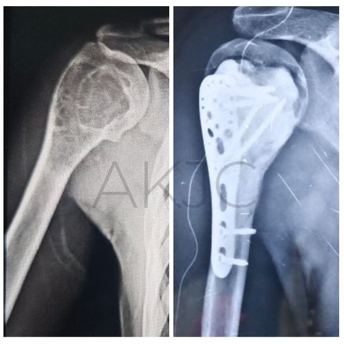
A 22-year-old patient from Mumbai presented with pain and swelling in the right shoulder, which had gradually worsened over time. patient had a history of fall 1 year back . Clinical examination, followed by imaging studies, revealed an expansile osteolytic lesion in the right proximal humerus — the upper part of the arm bone near the shoulder joint.
This raised concerns about the possibility of a bone tumor. To confirm the nature of the lesion, Dr. Anupam Khandelwal recommended a biopsy.
Diagnosis: Giant Cell Tumor (GCT)
The histopathology report from the biopsy confirmed the presence of a Giant Cell Tumor in the proximal humerus. GCTs are known for their locally aggressive behavior and can weaken bone structure, risking pathological fractures or joint dysfunction if left untreated.
Surgical Management: A Comprehensive Approach
Given the size, location, and aggressiveness of the lesion, Dr. Khandelwal planned a comprehensive surgical approach. The goals were clear: remove the tumor, prevent recurrence, restore structural integrity, and preserve joint function.
Here’s a step-by-step breakdown of the procedure:
1. Curettage
The tumor was first removed using ‘EXTENSIVE curettage’ , a procedure that involves scraping out the lesion from the bone.
2. Mechanical, Electrical, and Chemical Cauterization
To ensure thorough tumor eradication and minimize the risk of recurrence:
- Mechanical curettage was followed by
- Electrocautery to destroy residual tumor cells
- Chemical cauterization, using agents like phenol or hydrogen peroxide to reduce recurrence chances.
3. Pulse Lavage
The cavity was then cleaned using extensive pulse lavage, which involves high-pressure saline irrigation to clear out remaining debris and tumor fragments.
4. Bone Cement Filling and PHILOS Plate Fixation
Once the cavity was clean and stable:
- The defect was filled with bone cement (PMMA) to provide immediate structural support and allow for early mobilization.
- A titanium PHILOS (Proximal Humerus Internal Locking System) plate was used for rigid internal fixation to ensure joint stability and proper alignment.
Postoperative Recovery and Rehabilitation
One of the key highlights of Dr. Khandelwal’s treatment approach was early mobilization. Thanks to the stable fixation and support provided by bone cement and the PHILOS plate, the patient began guided physiotherapy shortly after surgery. This early movement is crucial to preserve shoulder mobility, minimize stiffness, and promote functional recovery.
Conclusion
This case demonstrates the importance of early diagnosis, multidisciplinary planning, and a structured surgical approach in managing complex bone tumors like Giant Cell Tumor. The patient, under the skilled care of Dr. Anupam Khandelwal, not only had the tumor successfully removed but also retained functional use of the shoulder — a remarkable outcome in such challenging cases.
About Dr. Anupam Khandelwal
Dr. Anupam Khandelwal is a renowned orthopedic surgeon known for his expertise in bone tumors, trauma surgeries, and joint reconstructions. His patient-centered approach and use of advanced surgical techniques make him one of the most trusted names in musculoskeletal care in India.