
Child Elbow Injury Care in Indore
Fractures Around the Elbow in Children
Pediatric elbow fractures need careful assessment because growth plates, cartilage and small ossification centres can make X-rays difficult to interpret.
A child with elbow swelling after a fall may have a supracondylar humerus fracture, lateral condyle fracture, medial epicondyle fracture, radial neck fracture, olecranon fracture or Monteggia-type injury.
Treatment depends on age, displacement, growth plate involvement, joint surface involvement, nerve and blood vessel status, and whether the fracture can be held safely after reduction.
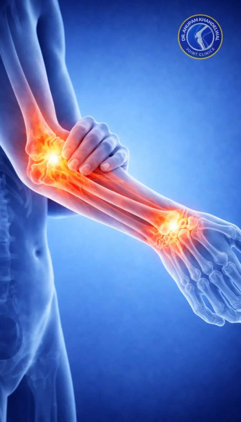
Common Elbow Fractures in Children
Pediatric elbow fracture diagnosis is based on injury mechanism, swelling pattern, X-ray signs and repeated neurovascular examination.
Supracondylar humerus fracture
Common after a fall on an outstretched hand. Displaced fractures may affect the brachial artery, median nerve, anterior interosseous nerve, radial nerve or ulnar nerve.
Lateral condyle fracture
Involves the growth plate and joint surface. Even moderate displacement can need fixation because late displacement, non-union or deformity can occur.
Medial epicondyle fracture
May occur with elbow dislocation. Fragment incarceration inside the joint, ulnar nerve symptoms or marked displacement changes management.
Radial neck fracture
Can reduce forearm rotation. Angulation, displacement and child age influence observation, closed reduction or fixation.
Olecranon fracture
Uncommon but important when there is extension weakness, displacement or associated radial head/neck injury.
Pulled elbow
A common non-fracture differential in young children with sudden refusal to use the arm, usually without swelling or deformity.
Urgent assessment
Do Not Delay Review if Circulation or Nerve Signs Are Present
A pale or cold hand, weak pulse, severe pain, increasing swelling, numbness, finger weakness, open injury or obvious deformity after a child elbow injury needs urgent orthopaedic assessment.
X-ray Assessment in Pediatric Elbow Fracture
Children have growth plates and ossification centres that can hide or mimic fracture lines. The whole elbow alignment must be reviewed, not only the obvious painful area.
Useful X-ray Clues
- AP and true lateral elbow views.
- Anterior humeral line and radiocapitellar alignment.
- Fat pad sign suggesting joint effusion or occult fracture.
- Comparison view in selected doubtful cases.
When Further Imaging Is Considered
- CT scan is rarely routine but can help with complex articular fractures.
- Ultrasound may help selected occult injuries depending on availability and expertise.
- Repeat X-ray is useful when initial images are doubtful but clinical suspicion remains high.
- MRI is reserved for selected cartilage, ligament or occult injury questions.
Treatment Options for Child Elbow Fractures
Treatment must protect fracture healing, growth plate function, joint surface alignment and elbow motion.
Non-Surgical Treatment
- Plaster or splint for stable, non-displaced fractures.
- Elevation, swelling care and pain control.
- Strict follow-up X-rays when late displacement is possible.
- Parent instructions for finger movement, swelling and circulation warning signs.
Reduction, Pinning or Fixation
- Closed reduction and percutaneous pinning for many displaced supracondylar fractures.
- Open reduction when closed reduction is unsafe, blocked or inadequate.
- Screw or K-wire fixation for selected lateral condyle or medial epicondyle fractures.
- Reduction of associated elbow dislocation before final fracture planning.
Follow-up, Growth and Motion After Pediatric Elbow Fracture
Follow-up is not only to remove plaster. It checks healing, alignment, elbow motion and late complications.
What Is Monitored
- Loss of reduction on repeat X-ray.
- Pin-site condition if K-wires are used.
- Return of elbow bending, straightening and forearm rotation.
- Carrying angle and growth-related alignment.
Possible Problems
- Cubitus varus or valgus deformity.
- Stiffness after immobilization or injury.
- Non-union risk in lateral condyle fracture if missed or undertreated.
- Nerve symptoms or vascular sequelae after severe injuries.
Book Consultation
Book consultation for a child elbow fracture
Book consultation if a child has elbow swelling, deformity, severe pain, reduced finger movement or X-ray evidence of fracture after a fall.
Bring AP and lateral X-rays, splint details and any hospital notes about pulse, sensation or attempted reduction.