
Adult Elbow Fracture Care in Indore
Fractures Around the Elbow
Adult elbow fractures can involve the radial head, olecranon, coronoid, distal humerus or a combined fracture-dislocation pattern.
The elbow is a small joint with three articulations: humeroulnar, radiocapitellar and proximal radioulnar. A few millimetres of articular step, gap, depression or malalignment can affect bending, straightening, forearm rotation and stability.
Treatment is decided after X-ray, clinical examination and often CT scan assessment. The surgeon decides between plaster, sling support, screw fixation, plate fixation, radial head reconstruction, radial head replacement or ligament repair according to the exact fracture pattern.
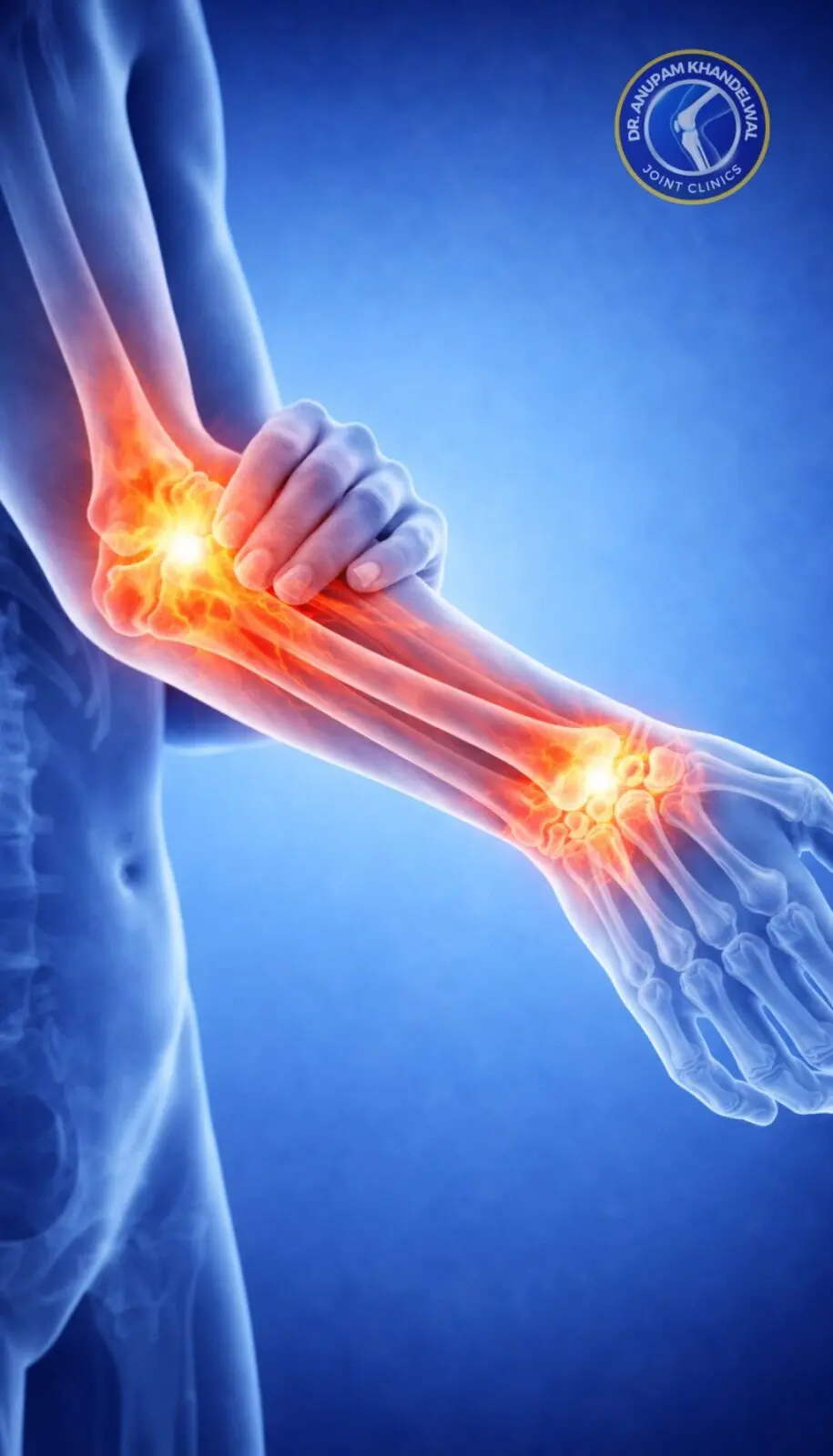
Important Adult Fractures Around the Elbow
Each fracture is assessed by location, displacement, joint involvement and whether the elbow remains stable through movement.
Radial head and neck fracture
Can block forearm rotation or destabilize the elbow when associated with ligament injury. Reconstructable fractures may be fixed; severely comminuted unstable patterns may need replacement.
Olecranon fracture
Involves the bony tip of the elbow where the triceps inserts. Displacement can weaken active elbow extension and disturb the trochlear notch joint surface.
Distal humerus fracture
May involve one or both columns, the capitellum, trochlea or the entire articular surface. CT scan helps define column fixation and joint reconstruction needs.
Coronoid fracture
A small coronoid fragment can be clinically important because the coronoid resists posterior elbow dislocation and contributes to stability.
Capitellum / trochlea fracture
Articular shear fractures can cause painful clicking, catching or blocked motion if the fragment is displaced.
Fracture-dislocation
Elbow dislocation combined with radial head, coronoid or ligament injury is treated as an instability problem, not only as a broken bone.
Urgent signs
Elbow Fractures Should Not Be Left Stiff or Unassessed
Rapid swelling, deformity, inability to rotate the forearm, numb fingers, weak pulse, open wound or increasing pain after an elbow injury needs urgent orthopaedic assessment.
X-ray and CT Scan Planning
X-ray confirms most elbow fractures. CT scan is added when the joint surface, fragment geometry or operative plan is unclear.
What X-rays Show
- AP view shows medial-lateral alignment, distal humerus columns and gross displacement.
- Lateral view shows olecranon alignment, coronoid involvement, radial head position and joint congruity.
- Fat pad sign may suggest occult fracture when the fracture line is subtle.
- Dislocation or subluxation is checked before and after reduction.
What CT Scan Adds
- Fragment size, number and orientation.
- Articular step, gap, depression and impaction.
- Coronoid subtype and radial head reconstructability.
- Column involvement in distal humerus fractures.
- Surgical approach and implant planning.
CT is especially useful before deciding reconstruction, replacement or complex fixation around the elbow.
When an Elbow Fracture May Be Treated Without Surgery
Non-surgical treatment is selected only when alignment, joint congruity and stability are acceptable.
Suitable Patterns May Include
- Minimally displaced radial head fracture without mechanical block.
- Stable non-displaced olecranon fracture with intact active extension.
- Small stable coronoid fracture without elbow subluxation.
- Occult or hairline fracture with preserved alignment.
Follow-up Priorities
- Short period of support in sling, splint or plaster as advised.
- Repeat X-rays to ensure displacement has not increased.
- Protected early movement when safe to reduce stiffness risk.
- Clear restriction on lifting, pushing and weight-bearing through the hand.
Surgical Options for Displaced or Unstable Elbow Fractures
Surgery aims to restore the articular surface, maintain stability and allow safe rehabilitation. The operation is selected by fracture anatomy, not by a fixed protocol.
Fixation and Reconstruction
- Headless compression screw fixation for suitable articular fragments.
- Low-profile radial head plate or mini-fragment plate when contour support is needed.
- Olecranon plate fixation or tension-band principle in selected patterns.
- Dual-column distal humerus plating for bicolumnar articular fractures.
When Replacement or Ligament Repair Is Considered
- Radial head replacement when fragments are too comminuted for reliable reconstruction.
- Lateral collateral ligament repair in unstable dislocation patterns.
- Coronoid fixation when the fragment contributes to instability.
- External fixation is reserved for selected severe instability or soft tissue situations.
Recovery and Stiffness Prevention After Elbow Fracture
The elbow becomes stiff quickly after injury. Rehabilitation timing depends on fracture stability, fixation quality, pain, swelling and soft tissue healing.
Early Recovery Goals
- Control swelling and pain without ignoring neurovascular warning signs.
- Protect the fracture or fixation during early healing.
- Start bending, straightening, pronation and supination when the surgeon permits.
- Avoid forceful massage or unapproved manipulation in fresh injuries.
Longer-Term Follow-up
- X-rays assess healing and implant position.
- Physiotherapy progresses from protected motion to strengthening.
- Return to lifting, driving, sport and work is individualized.
- Post-traumatic stiffness, non-union, malunion or arthritis are monitored.
Book Consultation
Book consultation for an adult elbow fracture
Book consultation if X-ray or CT scan shows radial head, olecranon, coronoid, distal humerus or fracture-dislocation injury around the elbow.
Early assessment helps decide whether safe mobilization, fixation, reconstruction, replacement or ligament stabilization is required.